Pathway Ureterolithiasis Doc
Batu dapat bermula dari pelvis renalis (batu ginjal atau nefrolithiasis) dan dapat berpindah ke ureter (ureterolithiasis), vesika urinaria (vesikolithiasis), atau uretra (uretrolithiasis). [1,2] Sebesar 80% batu pada urolithiasis terdiri dari kalsium oksalat atau fosfat.

Pathway of the Ureter Diagram Quizlet
Scribd adalah situs bacaan dan penerbitan sosial terbesar di dunia.

Ureter, bladder and urethra histology Osmosis
Batu ginjal atau nefrolitiasis merupakan suatu keadaan dimana terdapat satu atau lebih batu di dalam pelvis atau kaliks dari ginjal. Secara garis besar pembentukan batu ginjal dipengaruhi oleh faktor intrinstik dan ekstrinsik. Faktor intrinsik yaitu umur, jenis kelamin, dan keturunan.

Pathway Batu Ureter Fix
1. Nyeri berat di samping dan belakang, di bawah tulang rusuk 2. Nyeri yang menjalar ke perut bawah dan pangkal paha 3. Nyeri yang datang dalam gelombang dan berfluktuasi dalam intensitas 4. Nyeri saat buang air kecil 5. Urine yang berwarna merah muda, merah, atau cokelat 6. Urine keruh atau berbau tidak sedap

ASKEP BATU URETER PDF
Urolitiasis adalah proses terbentuknya batu (kalkuli) pada traktus urinarius. Diperkirakan 10% dari semua individu dapat menderita urolitiasis selama hidupnya, meskipun beberapa individu tidak menunjukkan gejala atau keluhan.

Surgical anatomy of the ureter Fröber 2007 BJU International Wiley Online Library
Pathway Batu Ureter | PDF. Scribd is the world's largest social reading and publishing site.
Urinary Tract System Ureter UrologyStone
Download Pathway Batu Ureter Type: PDF Date: June 2020 Size: 146.5KB Author: Gustiar Eighteenth This document was uploaded by user and they confirmed that they have the permission to share it. If you are author or own the copyright of this book, please report to us by using this DMCA report form. Report DMCA
Urinary System Structures
Renal stones are formed within the kidneys, and this is called nephrolithiasis. Urolithiasis is a condition that occurs when these stones exit the renal pelvis and move into the remainder of the urinary collecting system, which includes the ureters, bladder, and urethra. Many patients with urolithiasis can be managed with expectant management, analgesic, and anti-emetic medications; however.

Anatomy of the kidney and ureter Surgery Oxford International Edition
Ureteral obstruction surgery may be performed through one of these surgical approaches: Endoscopic surgery. This minimally invasive procedure involves passing a lighted scope through the urethra into the bladder and other parts of the urinary tract. The surgeon makes a cut into the damaged or blocked part of the ureter to widen the area and.

Endoscopic Management of Distal Ureteral Strictures Abdominal Key
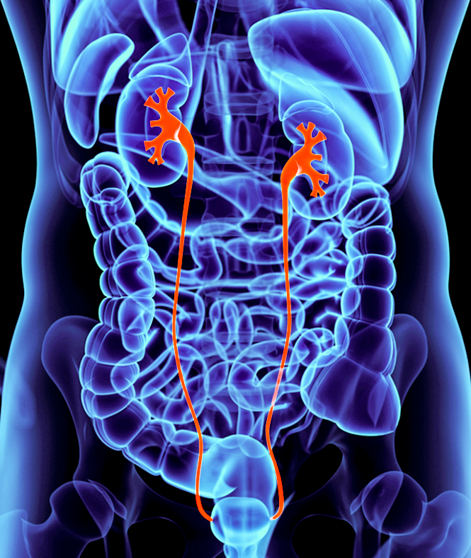
Batu ginjal atau dalam bahasa medis disebut sebagai nefrolitiasis adalah terdapatnya batu pada ginjal akibat kristalisasi berbagai mineral atau garam dalam urin. Selain di ginjal, batu juga dapat terbentuk di seluruh bagian saluran kemih (Gambar 1) seperti kandung kemih, ureter (saluran yang menghubungkan ginjal dan kandung kemih) dan uretra.

PPT Anatomy of he Urinary System PowerPoint Presentation, free download ID1898932
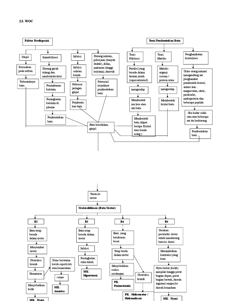
Dokumen tersebut membahas tentang pathway batu ureter, faktor penyebabnya, teori-teori pembentukan batu ginjal, tindakan bedah untuk mengeluarkan batu ginjal, dan komplikasi pasca operasi seperti infeksi, aliran balik urin, nyeri, serta dampaknya terhadap mobilitas dan perawatan diri pasien..
Ureter Anatomy QA
of 1 G. PATHWAY Genetik, Diet, Pekerjaan, Dehidrasi Batu Saluran Kemih Pre Operasi Intra Operasi Post Operasi Obstruksi Pemberian Anestesi Tirah Baring Saluran Kemih SAB Hambatan Rasa Aman Kaki tidak terasa Hambatan Aliran akibat anestesi Kemih Spasme batu saat Gangguan turun dari ureter Pergerakan Nyeri Akut Defisit Perawatan Hambatan
Ureters Function, Definition and Anatomy Human Anatomy Kenhub YouTube
Pathway Batu Ureter | PDF. Dokumen tersebut membahas tentang faktor-faktor penyebab terbentuknya batu ginjal, meliputi faktor intrinsik seperti hereditas dan umur, faktor ekstrinsik seperti asupan air dan diet, serta tahapan. by dichawening.

Ureter; Ureteres
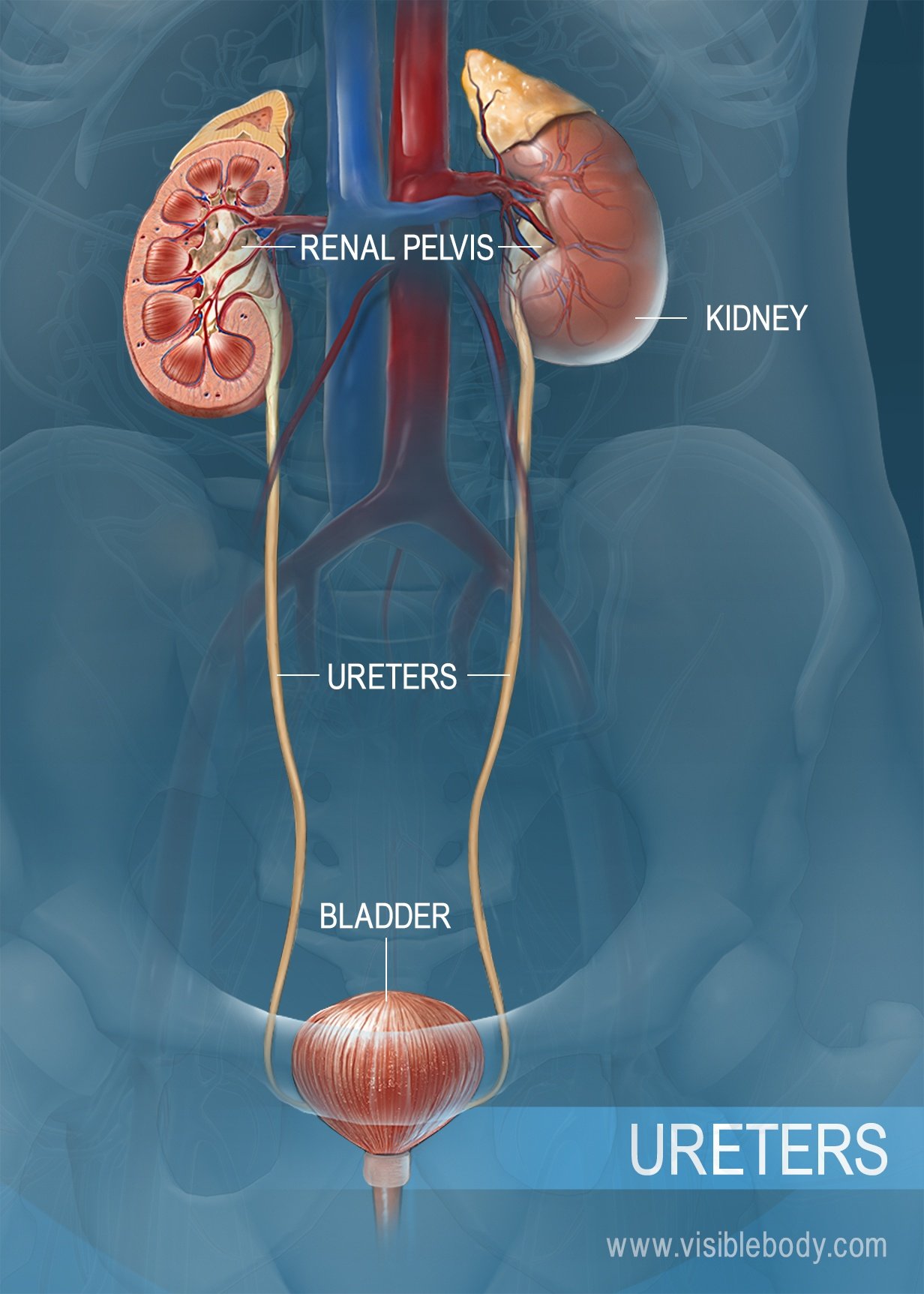
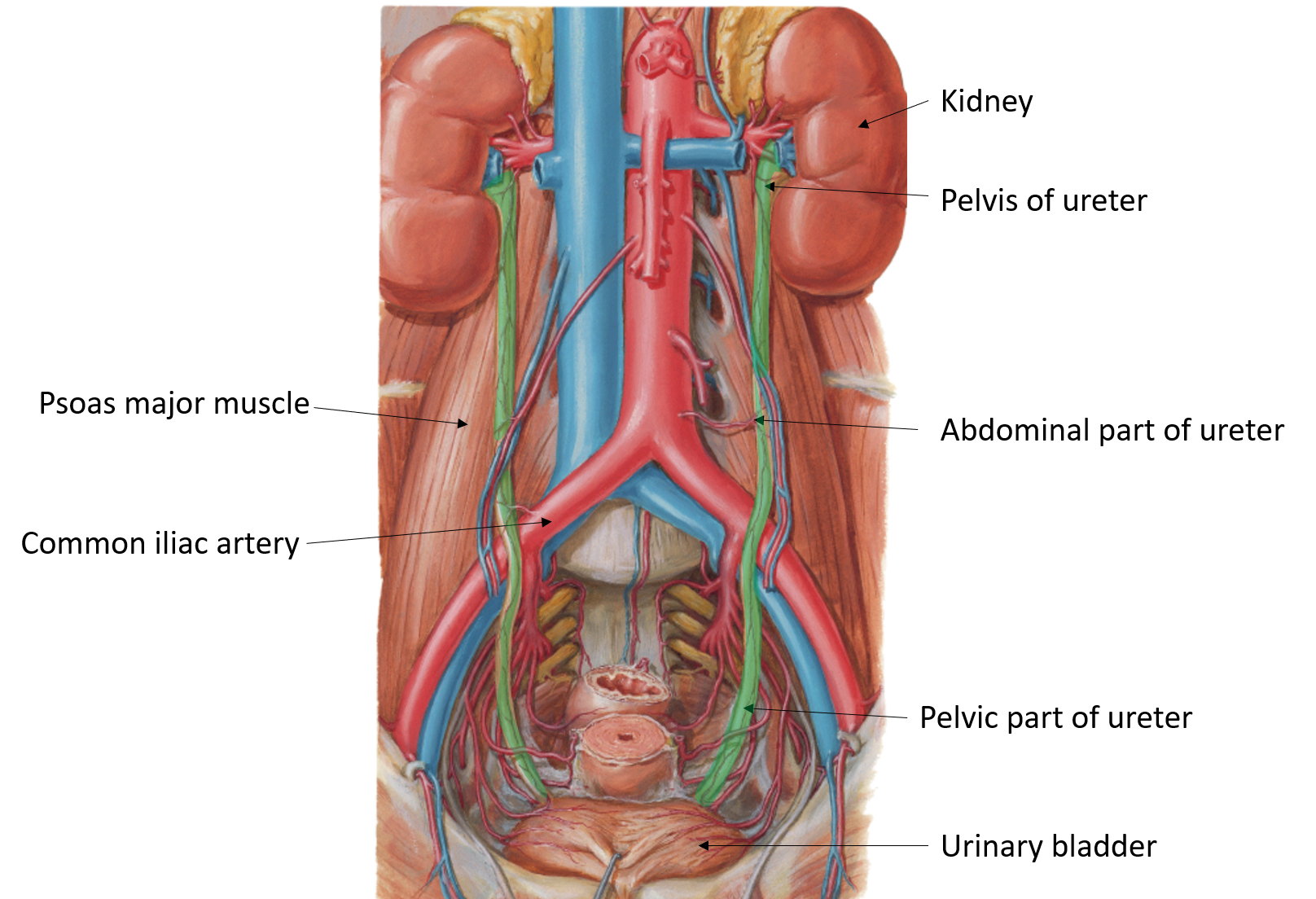
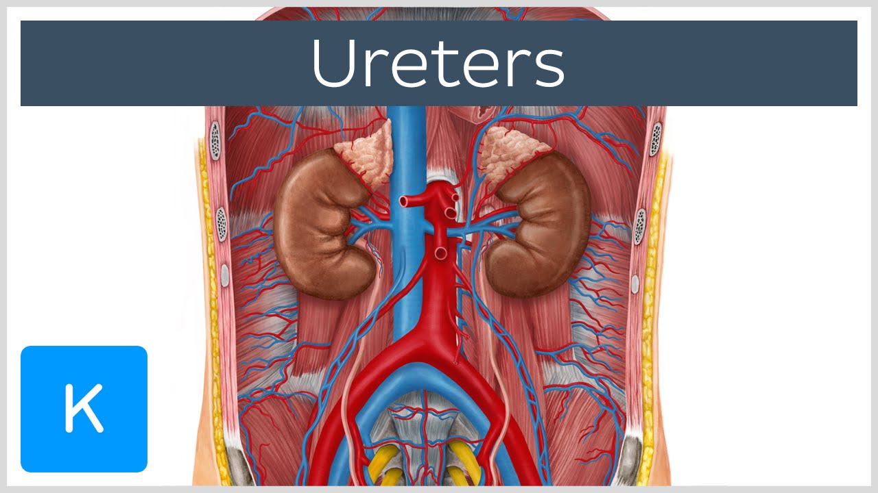
The Ureters. The ureters are two thick tubes which act to transport urine from the kidney to the bladder. They are approximately 25cm long and are situated bilaterally, with each ureter draining one kidney. In this article, we shall look at the anatomy of the ureters - their anatomical course, neurovascular supply and clinical correlations.

Genes and signaling pathways involved in ureter development. Scheme of... Download Scientific
The ureters are bilateral thin tubular structures with a 3 to 4 mm diameter that connect the kidneys to the urinary bladder (see Image. Posterior Thoracolumbar Surface Anatomy). These muscular tubes transport urine from the renal pelvis to the bladder. The ureter's muscular layers are responsible for the peristaltic activity that moves urine from the kidneys to the bladder.

The Ureters and Bladder Organization of the Urinary System The Urinary System Medical
Pathway Batu Ureter [klzz19jjz7lg].. Faktor intrinsic: Faktor idiopatik: Faktor ekstrinsik:-- Dehidrasi - ISK - Obstruksi saluran perkemihan - Asupan air - Diit - Pekerjaan Herediter Umur Jenis kelamin Defisiensi kadar magnesium, sifrat prifosfor, mukoprotein dan peptid Mual muntah Resiko kristalisasi mineral Penumpukan kristal Risiko tinggi kekurangan Pengendapan batu saluran kemih volume.