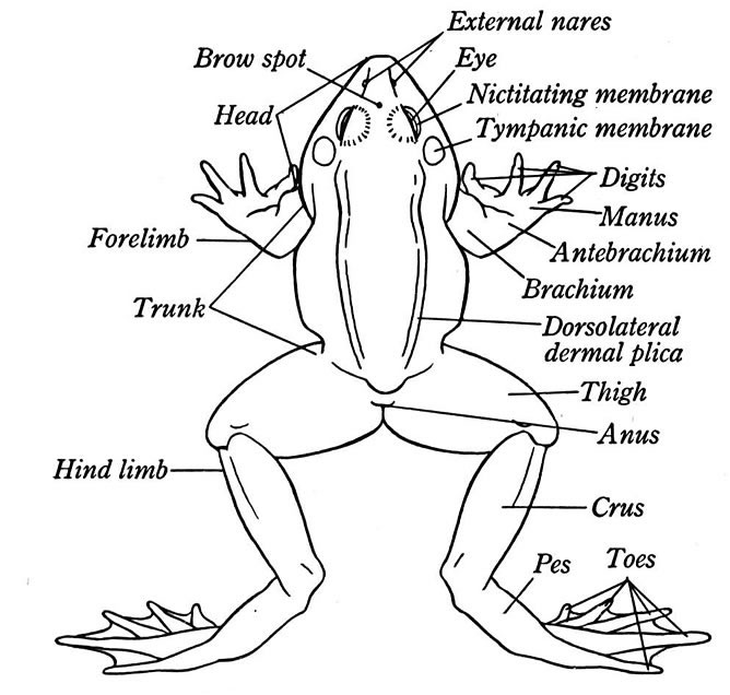
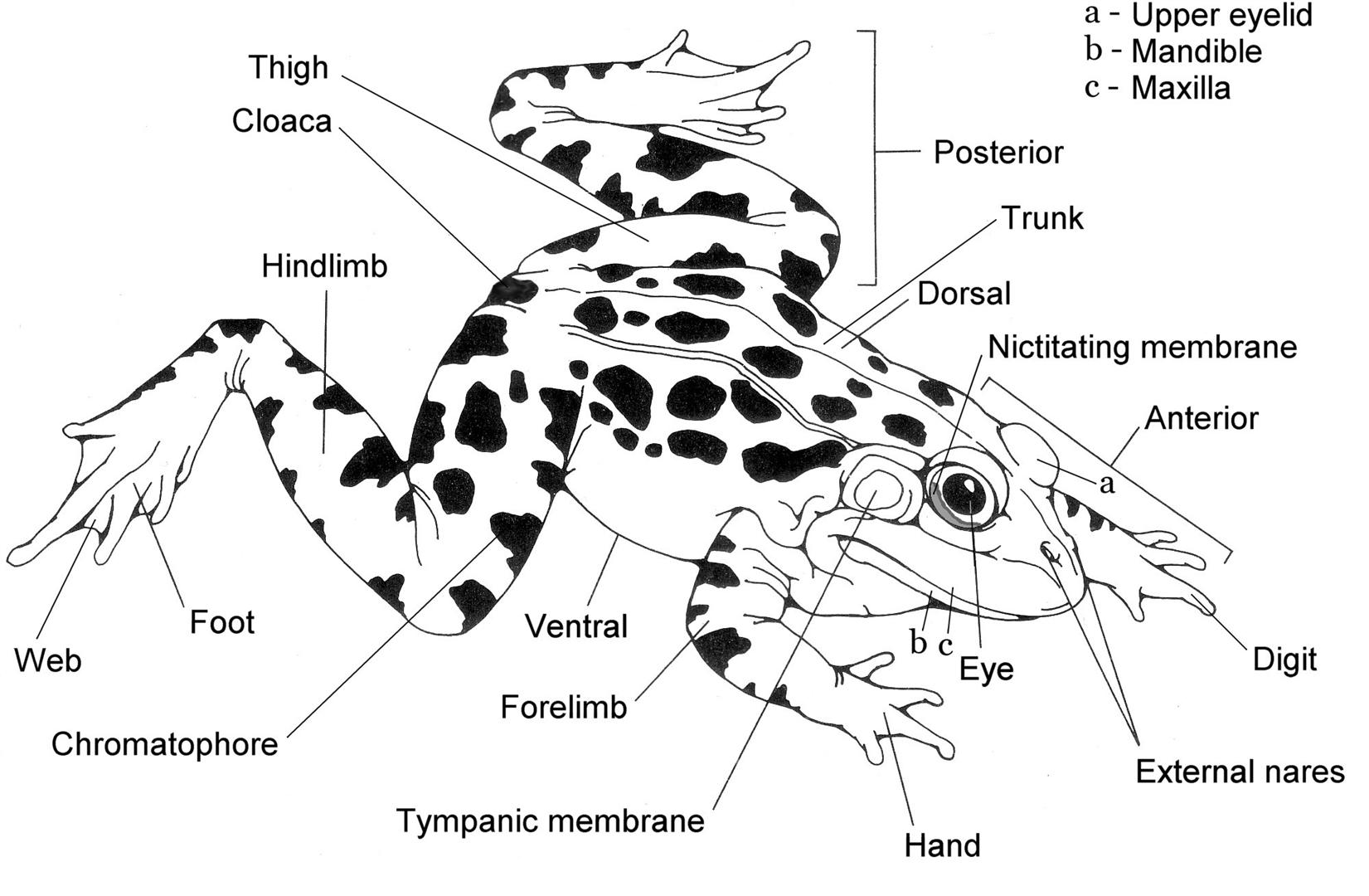
External Anatomy Of A Frog Diagram Of A Frog
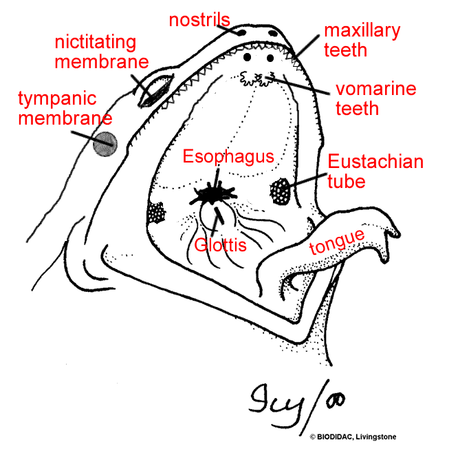
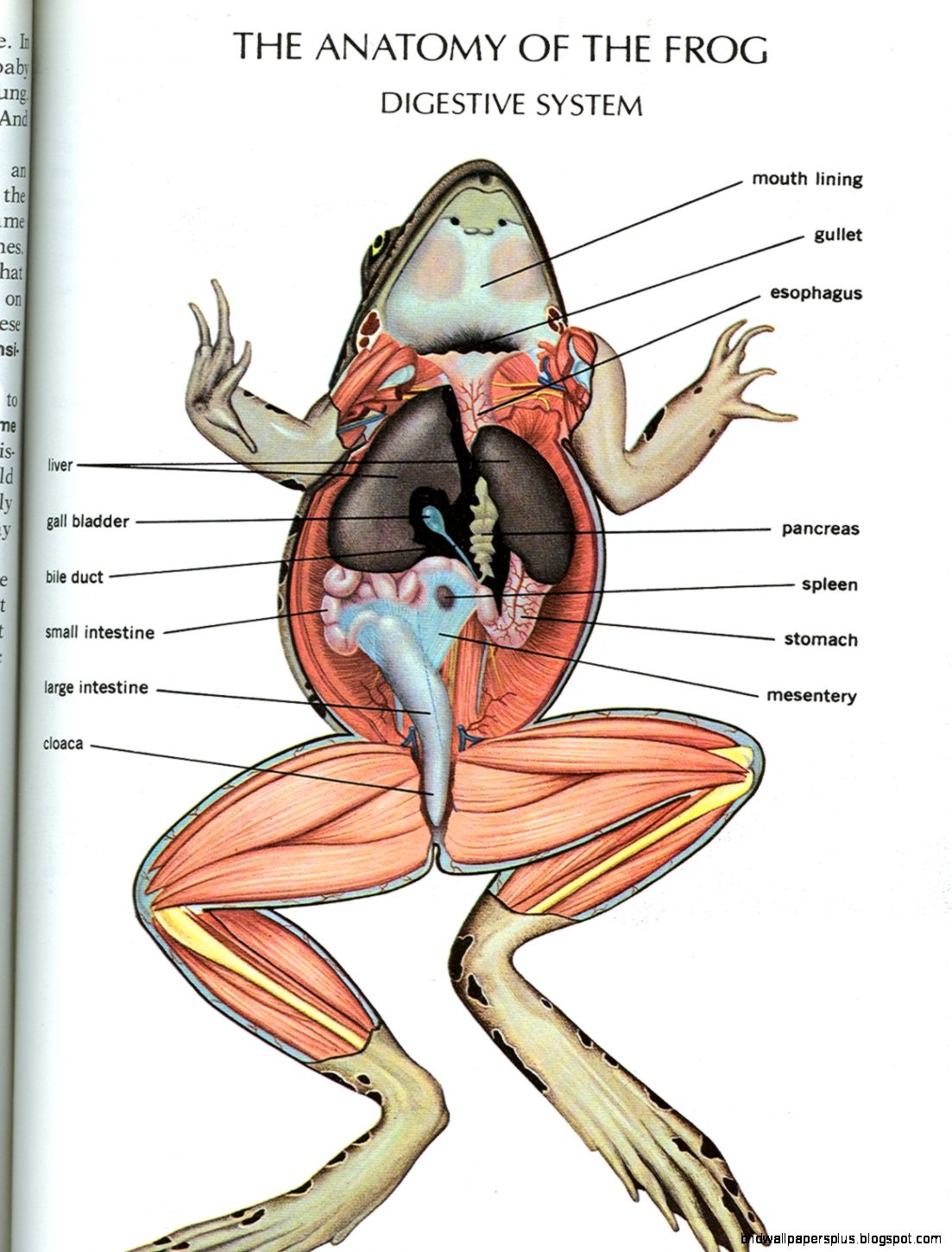
The transparent eyelid is called the nictitating membrane. You will also find the frog's stomach when you do a frog dissection. You will be able to open up the frog's stomach and see what the frog has eaten recently. You might find the wing of a fly or even a whole insect inside the stomach.
Frog anatomy labeled scheme Royalty Free Vector Image
Dissection Instructions. Place the frog in the dissecting pan ventral side up. Use scissors to lift the abdominal muscles away from the body cavity. Cut along the midline of the body to the forelimbs. Make transverse (horizontal) cuts near the arms and legs. Life the flaps of the body wall and pin back.
Anatomy of the Frog Ms. McGee's Science Class
Very few species on Earth have this ability. Frogs have been found as far back as 250M years ago. As of today, there are over 7,200 identified frog species worldwide. Most of them have similar internal anatomy, regardless of their size. I know you probably have an adult frog on the dissection table so we will get to that in a few seconds.
Frog Dissection Diagram and Labeling
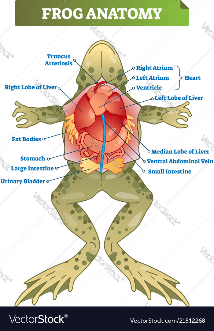
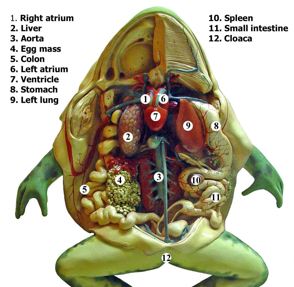
The Organs of the Abdominal Cavity Peritoneum: Spiderweb like membrane that covers organs Stomach: First site of chemical digestion, breaks down food Pyloric Sphincter - valve between stomach and small intestine Liver: Makes bile (aids in digestion) Gall bladder: Stores bile Esophagus: Tube that leads to the stomach
Biology 2
A diagram of the skeleton of a frog. Looking at how a Frogs bone structure is made up and what bones contribute to everyday life.. Skeletal anatomy of a Frog. Skeletal anatomy of a frog. Search. Most Popular Animals. Zebras; Aquatic Warbler; Atlantic Dolphins; Trapdoor Spider; Giraffe; Meerkats; Timber Wolf;
Frog Anatomy HD Wallpapers Plus
Frog Internal Anatomy - Dissection Guide. Lay the frog on its back, spread out its limbs, and pin them to the tray. Use forceps to lift the skin between the hind legs and make a small incision with a scalpel. Continue the cut up the center of the frog's body with scissors, being careful to cut through the skin only.
Frog Anatomy Overview 1 Carlson Stock Art
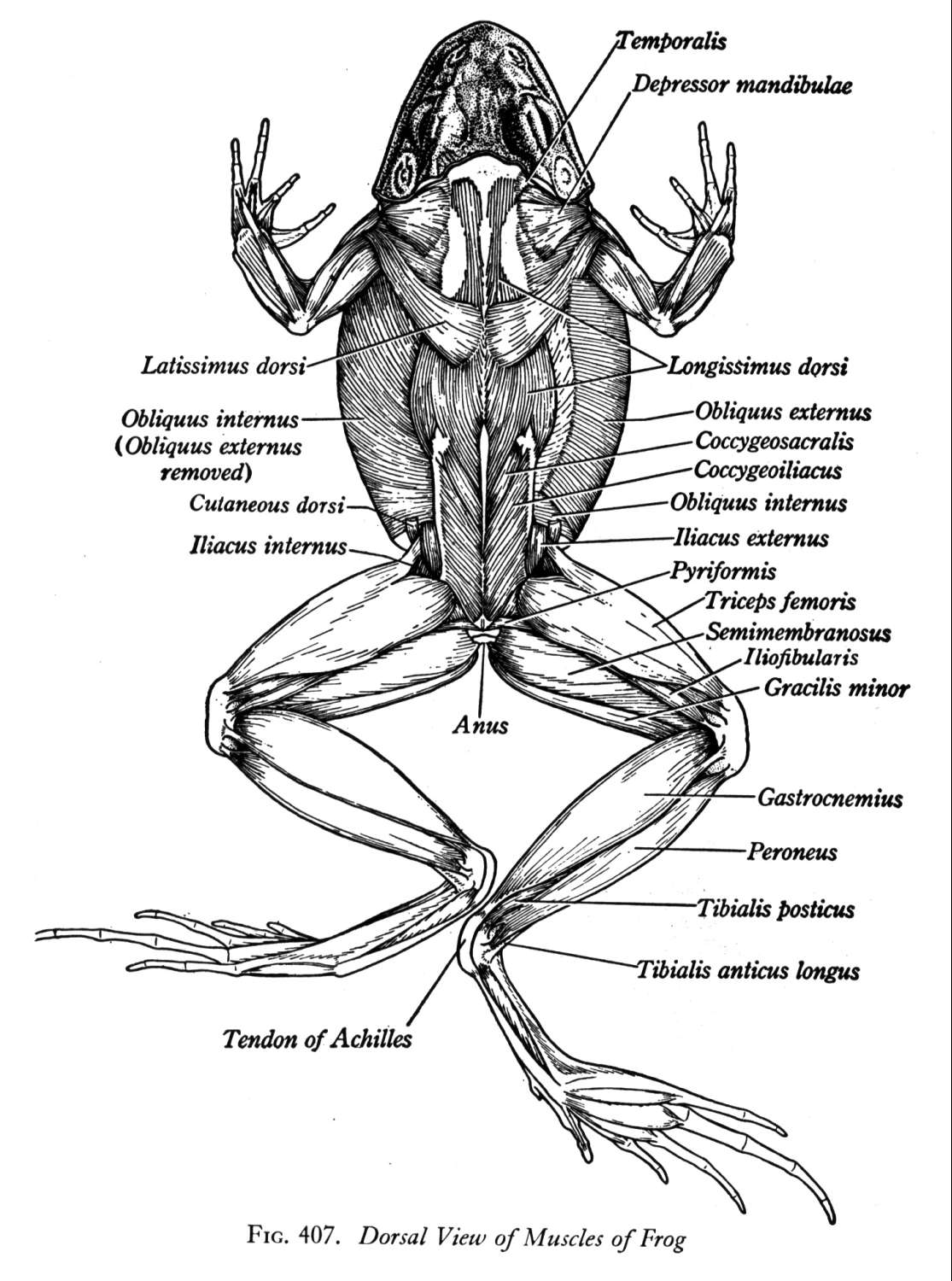
19 - Anatomy of the Frog. In this lab exercise, you were introduced to vertebrate anatomy through a frog dissection. Consult your lab manual for the organs that you will need to recognize on the frog dissection and model and know their functions. You will be expected to be able to identify the muscles of the hind limb and know their actions. In.
Frog Dissection Diagram and Labeling
When a flexor of a leg or other body part contracts, that part is bent. When the extensor of that body part contracts, the part straightens. Objectives: • Describe the appearance of various organs found in the frog.

Frog Anatomy Coloring Worksheet Biology LibreTexts
Frog External Anatomy - legs, eyes, mouth structures. Frog Dissection - major organs of the digestive, urogenital, and circulatory and respiratory systems. Frog Brain and Bones - remove the frog's brain, expose the bones of the lower leg. Frog Dissection Crossword - review terms and procedures. Observe a Living Frog - non dissection.
External Anatomy Of A Frog Anatomical Charts & Posters
A 3D Visual Guide to Frog Anatomy Posted on 11/18/22 by Sarah Boudreau There's a reason frog dissection is the quintessential biology lab: examining frog anatomy teaches students about how organ systems function in complex organisms, drawing similarities between frog anatomy and their own.

Free and Printable Frog Diagram 101 Diagrams
The style of citing shown here is from the MLA Style Citations (Modern Language Association). When citing a WEBSITE the general format is as follows. Author Last Name, First Name (s). "Title: Subtitle of Part of Web Page, if appropriate." Title: Subtitle: Section of Page if appropriate. Sponsoring/Publishing Agency, If Given.
All about frogs and toads Wildlife
Table of Contents Frog Skeleton Refer to the interactive diagram above to learn where each part is located. Maxilla - Forms the upper jawbone Atlast - The top part of a backbone Suprascapula - Shoulder blade Vertebrae - Individual bones that form the spine Sacral Vertebra - A bone below the last vertebra, positioned between the hips
Frog Anatomy Chart Flinn Scientific
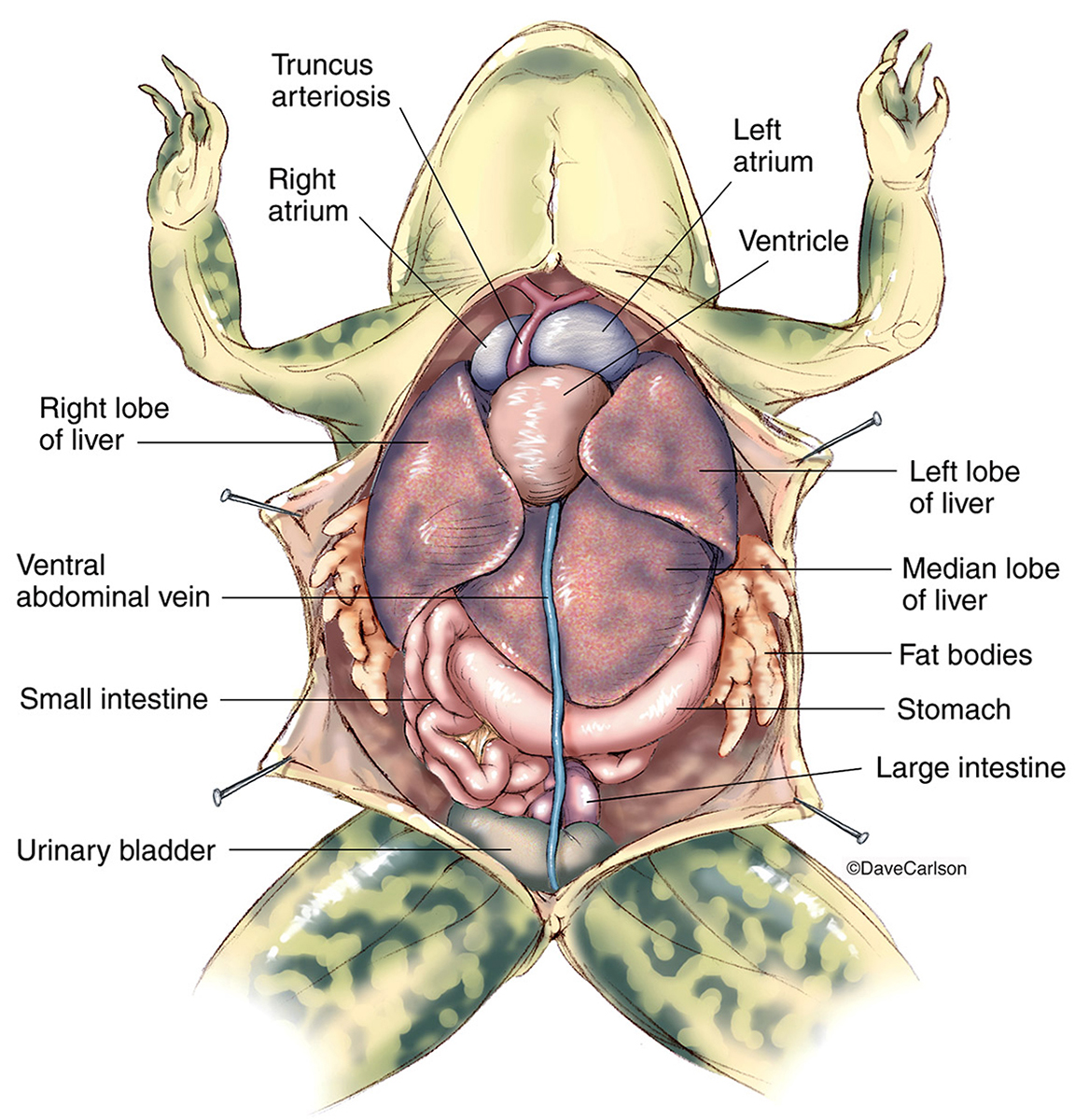
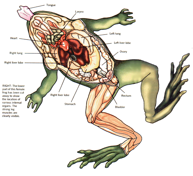
In the abdominal cavity, you can see the liver, stomach, intestines, kidneys, pancreas, fat bodies, testes (male), or ovaries (female). What is the external anatomy of a frog? The external.
11 Best Images of Frog Dissection Worksheet Frog Dissection Labeling
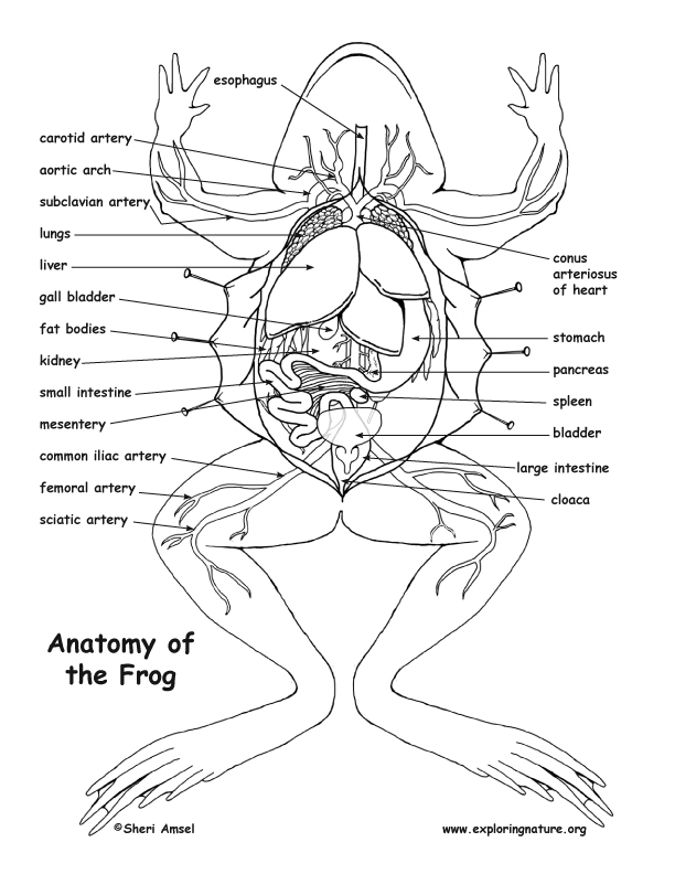
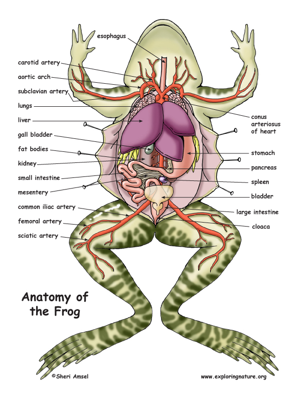
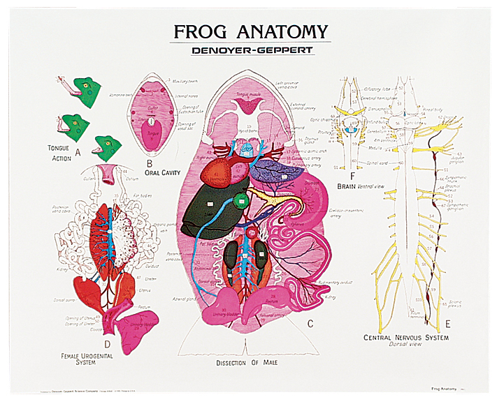
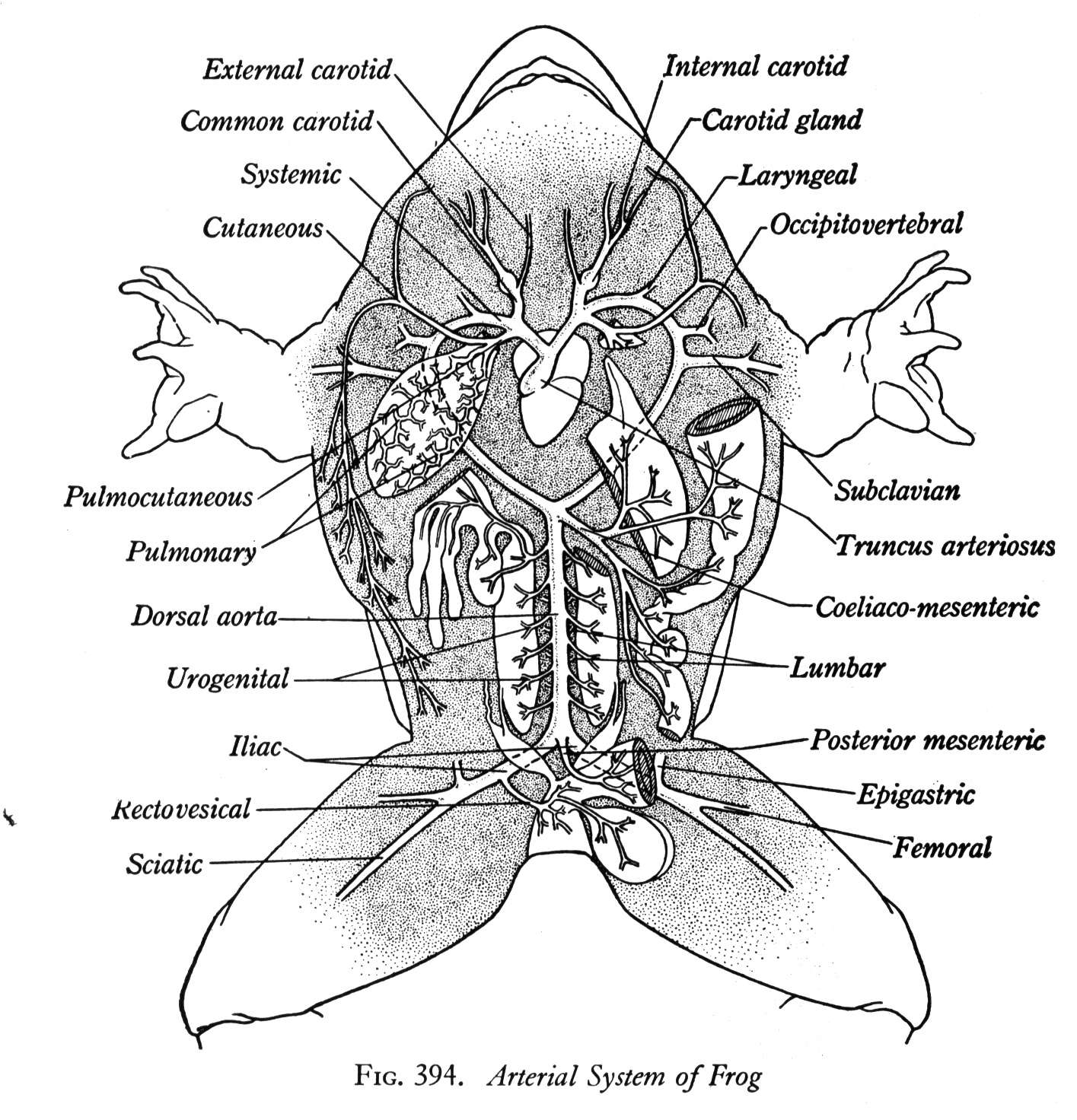
cloaca Label the Anatomy of the Frog esophagus carotid artery aortic arch subclavian artery lungs liver gall bladder fat bodies kidney small intestine mesentery conus arteriosus of heart stomach pancreas spleen bladder common iliac artery femoral artery sciatic artery large intestine cloaca Anatomy of the Frog
Anatomy of the Frog Ms. McGee's Science Class
Frogs' teeth are not used for chewing! Instead, their special vomerine teeth (shown as 'premaxillary teeth" on the frog anatomy app) are used to hold prey in place before swallowing. The vomerine teeth are notably pointy and appear in pairs of tiny clusters at the top front of the mouth. Elisabeth Ormandy, 2020. 18

Internal anatomy of the frog Animal Anatomy Pinterest Frogs
Below is an easy and well labelled diagram of frog ( Rana tigrina) for your better understanding. Anatomy The body plan of frogs consists of well-developed structures which help them in their physiological activities.