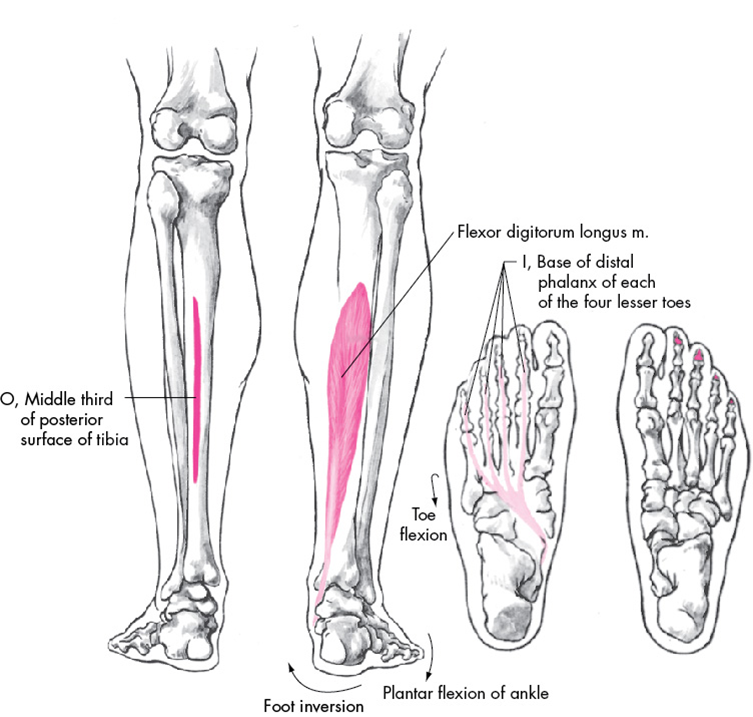
M Tibialis Posterior
⭐ Tibialis Posterior Muscle Anatomy ⭐ 💪 Origin: Posterior surface of tibia, posterior surface of fibula, posterior interosseous membrane. 💪 Insertion: Navicular bone tubercle,.

Tibialis Posterior Origin And Insertion
Tibialis posterior . Origin. Posterior aspect of interosseous membrane, superior 2/3 of medial posterior surface of fibula, superior aspect of posterior surface of tibia, and from intermuscular septum between muscles of posterior compartment and deep transverse septum. Insertion. Splits into two slips after passing inferior to plantar.
Tibialis Posterior Muscle Attachments, Actions & Innervation GetBodySmart
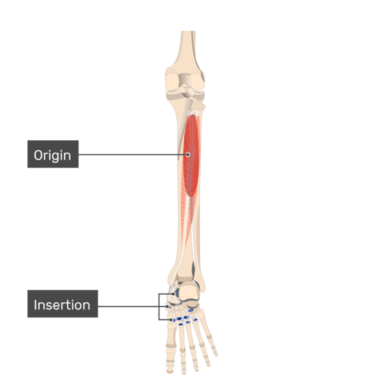
Definition Origin: Tibia, fibula Insertion: Navicular, medial cuneiform Artery: Posterior tibial artery Nerve: Tibial nerve Action: Inversion of the foot, plantar flexion of the foot at the ankle Antagonist: Tibialis anterior muscle Description:

tib post origin and insertion Google Search Muscle Origins and Insertions Pinterest
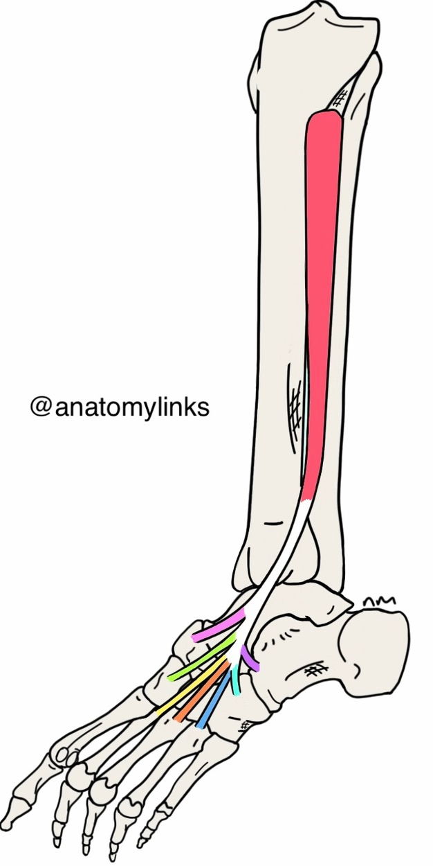
The tibialis posterior muscle originates from the interosseous membrane, while the posterior surface of the adjoining parts of the tibia, fibula, and muscle belly becomes the tibialis posterior tendon (TPT) in the distal third of the calf.

insertion/origin of tibialis posterior and flexors Diagram Quizlet
Soleal line: oblique line located on the posterior tibia and serves as the origin for the soleus, flexor digitorum longus, and tibialis posterior muscles. Serves as the origin or insertion point of many muscles including tibialis anterior, extensor digitorum longus, soleus, tibialis posterior, flexor digitorum longus, sartorius, gracilis, quadriceps femoris, semimembranosus, semitendinosus.

Tibialis Posterior Muscle Calf and Foot Pain The Wellness Digest
1 2 3 4 Attachments of Tibialis Posterior Muscle: Origin & Insertion Origin: (proximal attachments): a. Lateral portion of posterior, proximal tibia. b. Interosseous membrane. c. Medial portion of posterior, proximal half of fibula. Insertion: (distal attachments):
Tibialis Posterior Muscle Dr. Justin Dean
Tibialis posterior is attached between the bones of the leg and the foot. The muscle consists of two parts close to its origin; medial and lateral. The medial portion arises from the upper two-thirds of the posterior surface of tibia, inferior to the soleal line, and from the posterior surface of interosseous membrane of leg.

Image result for tibialis posterior origin and insertion Musculoskeletal system, Muscle and
Description The Tibialis Posterior is located deep in the posterior compartment of the lower leg and situated between the Flexor Digitorium Longus and the Flexor Hallucis Longus. It is a key stabilising muscle supporting the medial arch of the foot. Origin The origin of the muscle is [1] : Proximal postero-lateral aspect of the tibia.

Tibialis Posterior Insertion
The described anatomical insertion of the TPT may vary according to the geographical origin, ethnicity, and the number of examined specimens. Nevertheless, the sample size of 41dissected feet constitutes a good sample size.. In conclusion, this study adds to current knowledge on the anatomical insertion of the tibialis posterior tendon. The.
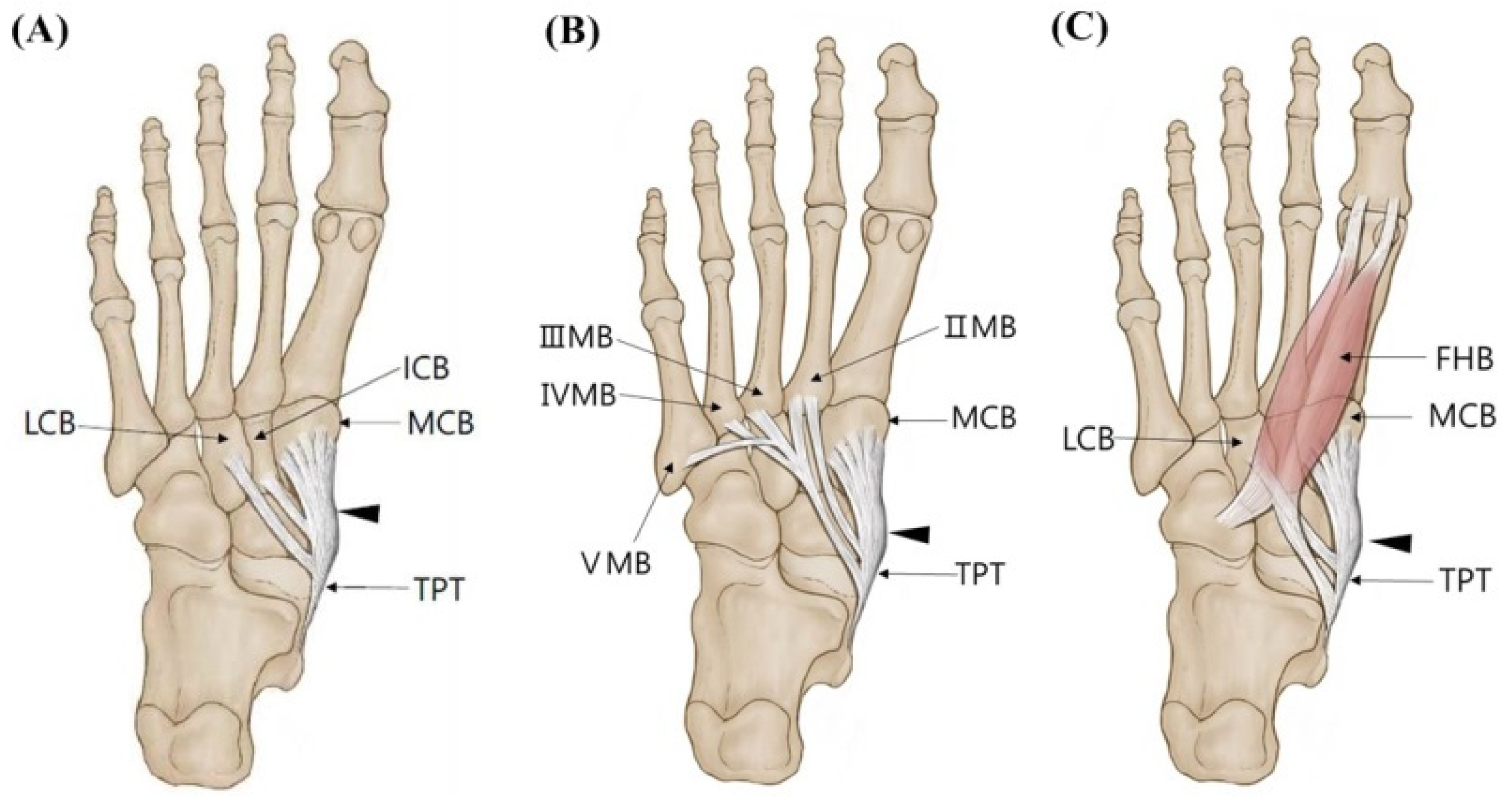
Diagnostics Free FullText A New Anatomical Classification for Tibialis Posterior Tendon
The tibialis posterior muscle, originating from the proximal tibia and fibula, passes distally with a broad insertion on the plantar aspect of the navicular, cuneiform, cuboid, and metatarsal bases and normally functions to invert the subtalar joint and to adduct the forefoot.

Tibialis Posterior Origin and Insertion Anatomy and physiology, Anatomy, Med school
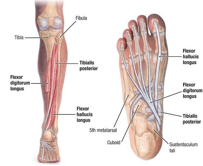
Origin: The posterior tibialis originates from the superior two-thirds of the medial posterior surface of the tibia. Insertion: The tendon of the posterior tibialis courses distally, bifurcating at the calcaneonavicular ligament, to insert on the tuberosity of the navicular bone (superficial slip) and the plantar surfaces of the second, third, and fourth metatarsals (deep slip).
Tibialis posterior Origins, insertions and actions Kenhub
The tibialis posterior muscle is one of the small muscles of the deep posterior compartment of the leg. Summary origin: upper half of posterior shaft of tibia and upper half of fibula between medial crest and interosseous border, and adjacent interosseous membrane. insertion: navicular and medial cuneiform

Tibialis Posterior Anatomy Study Origin, Insertion, Action, Innervation and Blood Supply The
The tibialis posterior muscle (TPM) is the deepest muscle of the deep posterior compartment of the lower leg. Its long muscle belly arises from the posterior aspect of the interosseous membrane and superior two-thirds of the posterior and medial surface of the fibula, and the superior aspect of the proximal tibia.
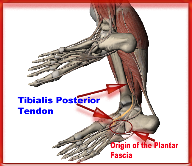
Endurance Athlete Consulting by Senska Physical Therapy Plantar Fasciitis or Tibialis Posterior
The tibialis posterior muscle originates from the: - lateral aspect of the area of the posterior surface of the tibia that is located inferior to the soleal line; - medial aspect of the proximal two thirds of the posterior surface of the fibula; - posterior aspect of the adjacent interosseous membrane of leg; - adjacent intermuscular septa.
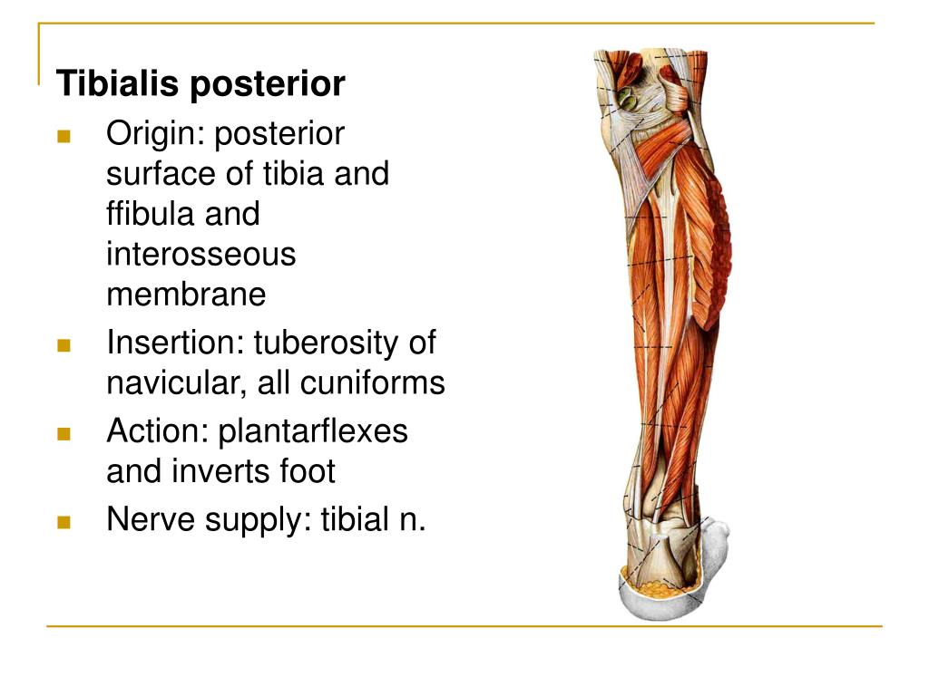
Tibialis Posterior Origin, Insertion, Anatomy and Function
The tibialis posterior muscle is the most central of all the leg muscles, and is located in the deep posterior compartment of the leg. It is the key stabilizing muscle of the lower leg . Posterior Tibial Tendonitis Posterior Tibial Tendonitis is a condition that predominantly affects runners and active individuals.
PPT The lower limb(1) PowerPoint Presentation, free download ID931555
Introduction. The tibialis posterior (TP) muscle has a vital role during gait; via multiple insertion points into the tarsal bones it acts as the primary dynamic stabiliser of the rearfoot and medial longitudinal arch (MLA) [1,2].The significance of TP function is evident when the muscle and tendon are dysfunctional, whereby stability of the foot is compromised and is associated with a.