
Cureus Simultaneous Bilateral Spontaneous Pneumothorax A Rare Complication of
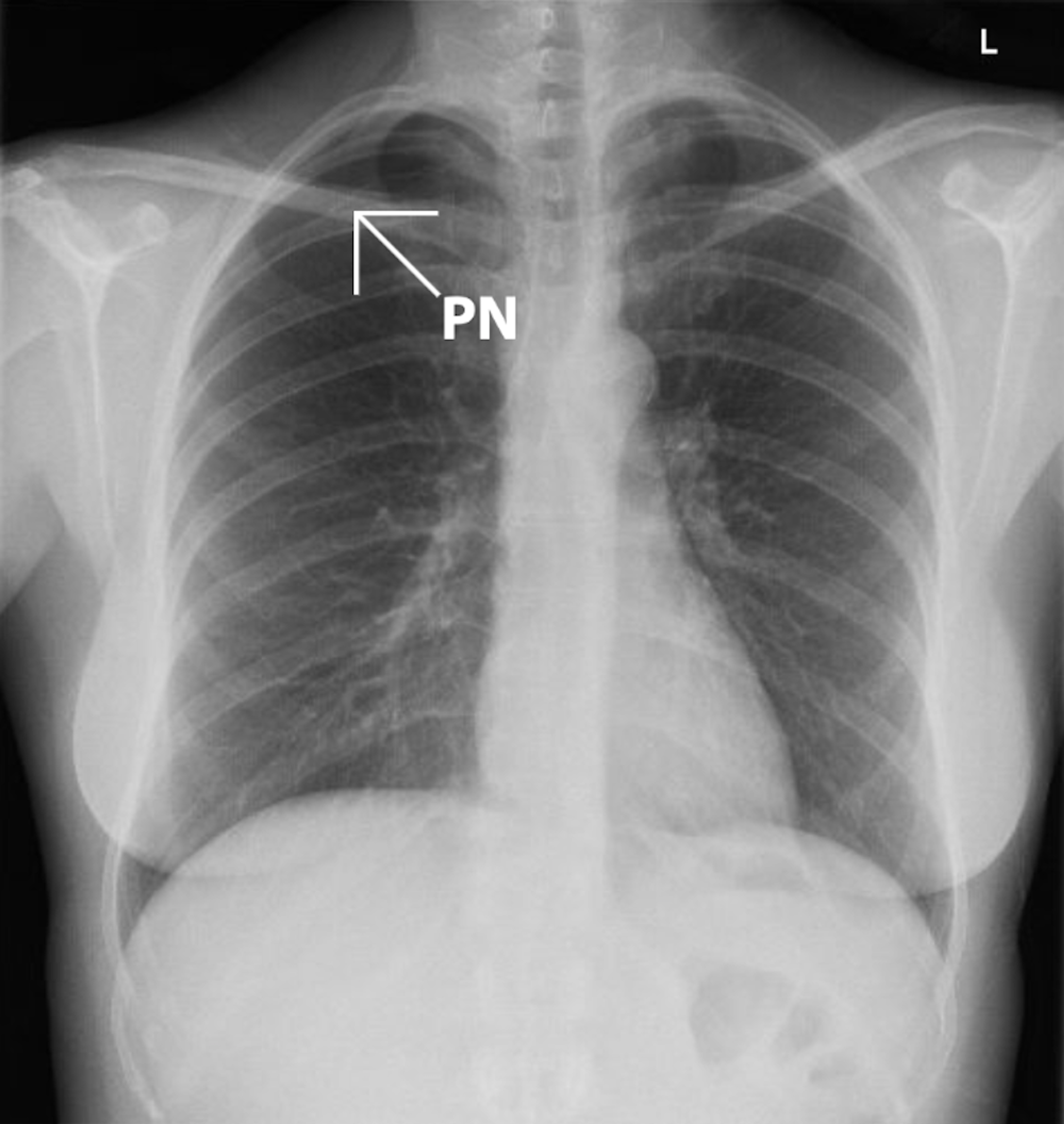
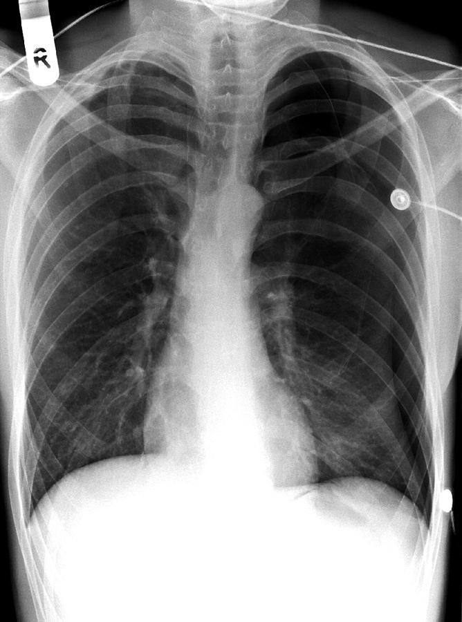
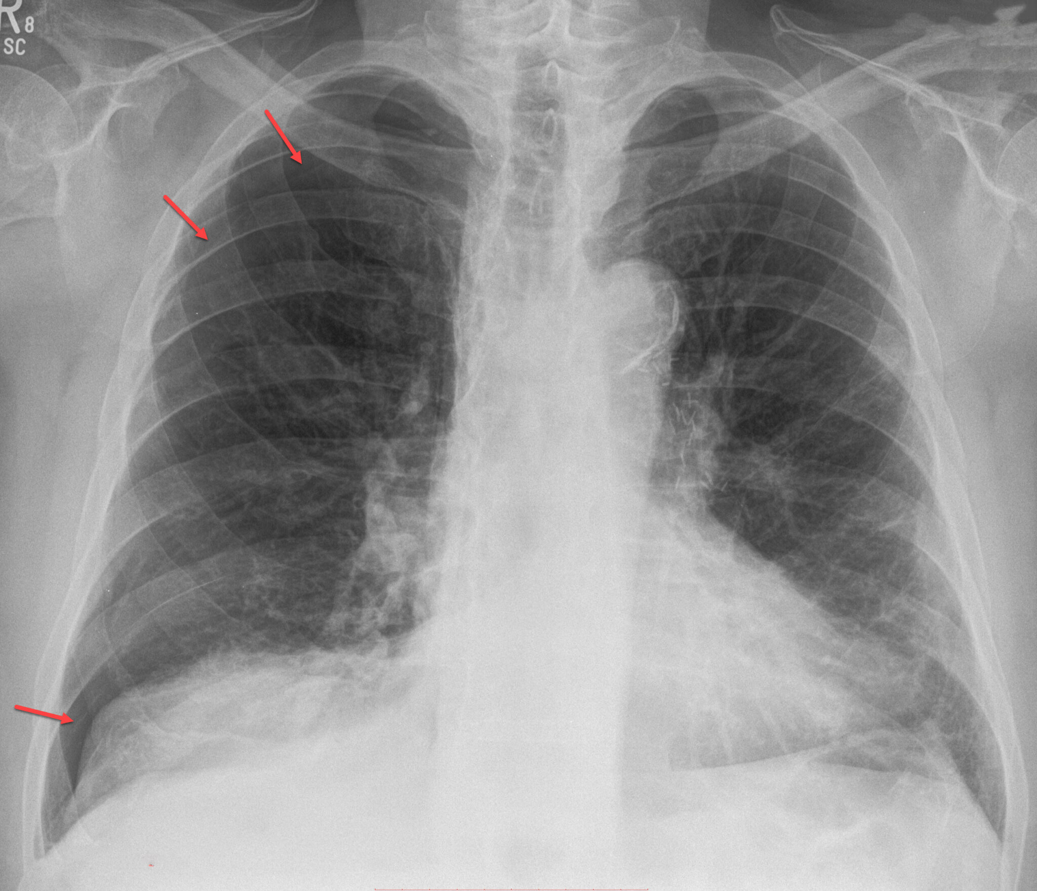
Chest X-ray (Figures 2 and 3): visible rim between the lung margin and chest wall, with an absence of lung markings. The size of the pneumothorax is measured at the level of the hilum: >2cm is classified as a "large" pneumothorax. 17; CT chest (Figure 4): may be used to identify small pneumothoraces missed by chest X-ray.
Cureus Pneumothorax Following Acupuncture
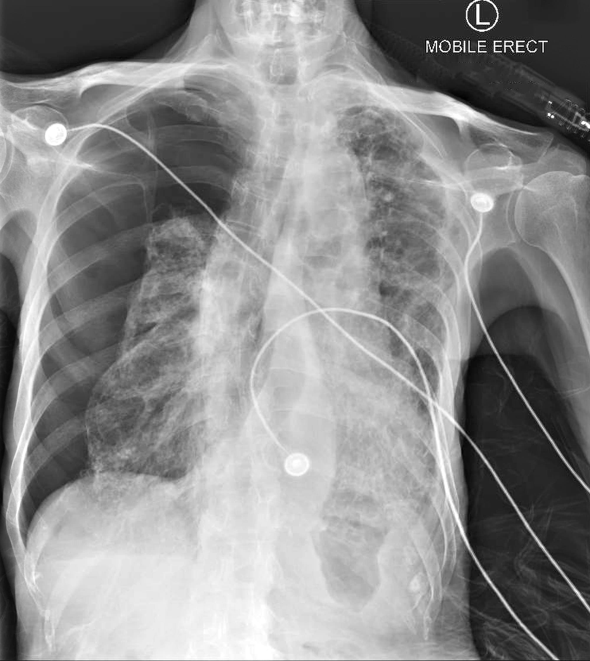
A non-tension pneumothorax is properly called a simple pneumothorax. Clinical presentation. Presentation is variable and may initially have no symptoms. With time severe dyspnea, tachycardia and hypotension occur. Distended neck veins and tracheal deviation are also often present. Eventually, impaired venous return results in cardiac arrest and.
Pneumothorax, Xray Photograph by Science Photo Library Pixels
In contrast, tension pneumothorax is a medical emergency and may be treated before imaging - especially if there is severe hypoxia, very low blood pressure, or an impaired level of consciousness. In tension pneumothorax, X-rays are sometimes required if there is doubt about the anatomical location of the pneumothorax. Chest X-ray
Pneumothorax Chest XRay MedSchool
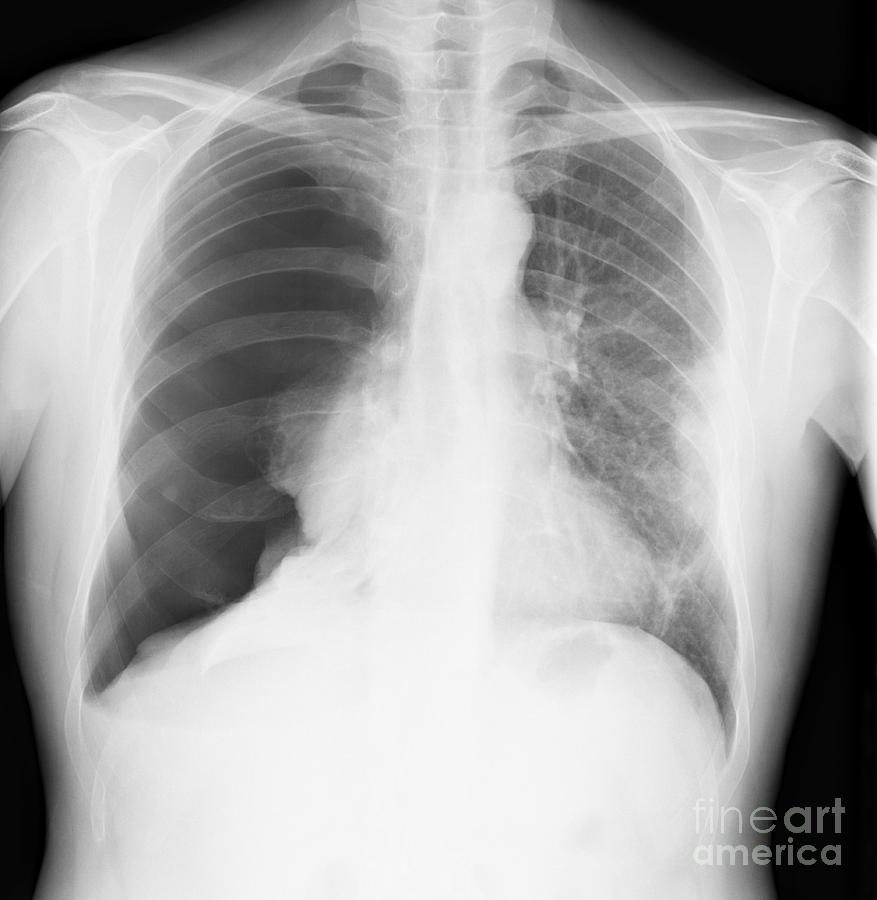
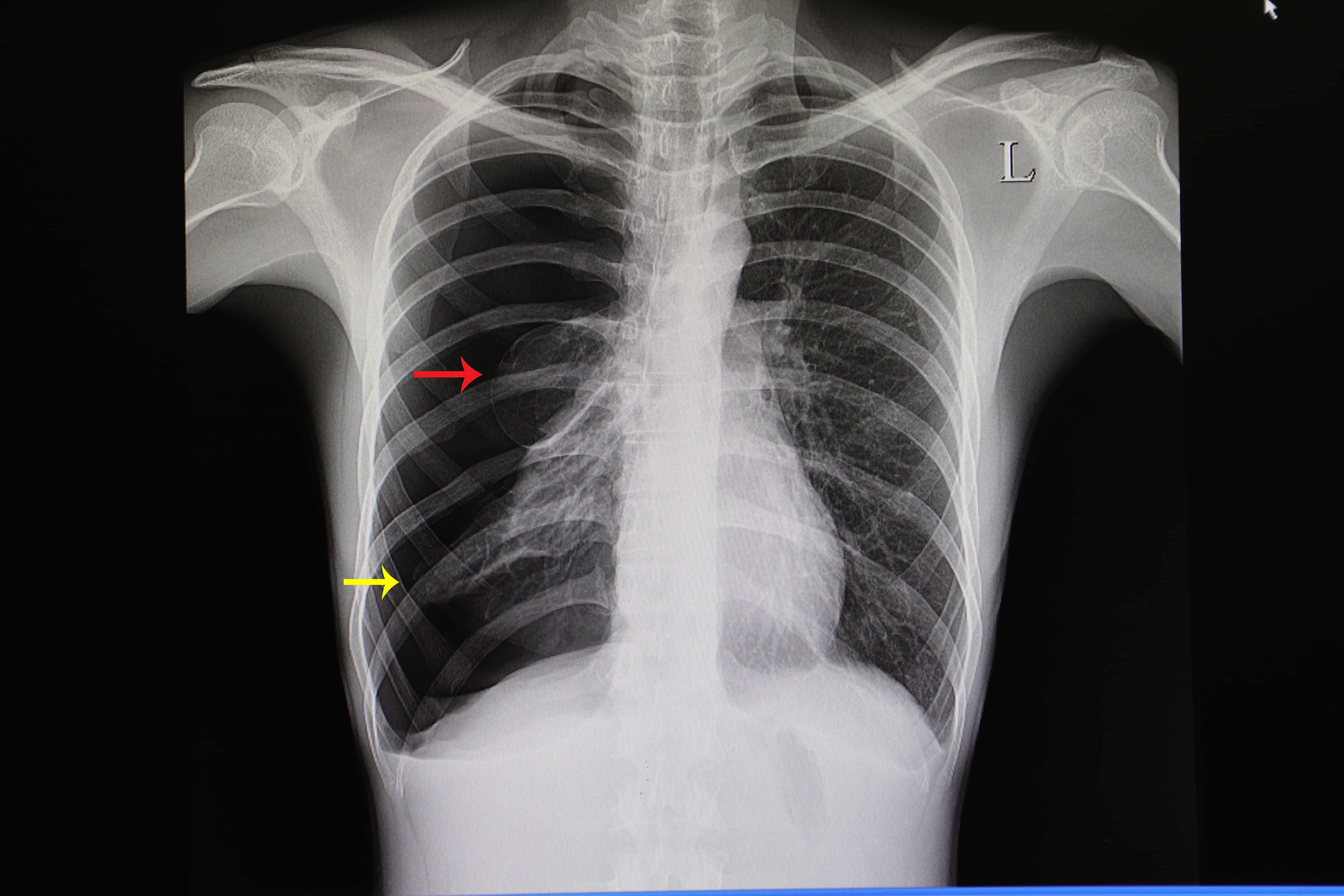
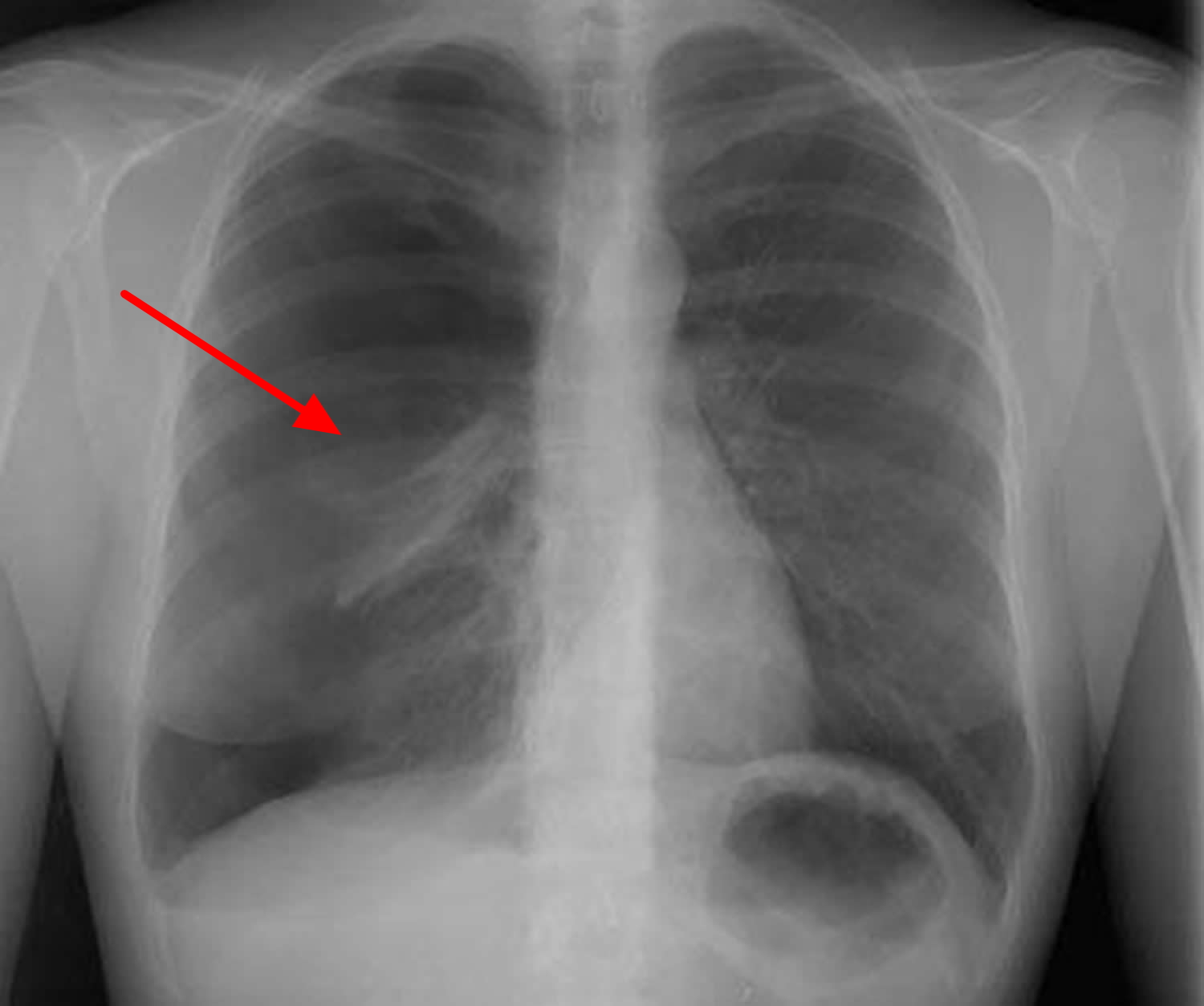
A pneumothorax is, when looked for, usually easily appreciated on erect chest radiographs. Typically they demonstrate: visible visceral pleural edge is seen as a very thin, sharp white line. no lung markings are seen peripheral to this line. peripheral space is radiolucent compared to the adjacent lung.
Pneumothorax, Xray Photograph by Science Photo Library
Pneumothorax is an urgent situation that has to be treated immediately upon diagnosis. Pneumothorax is divided to primary and secondary.. Diagnosis of a pneumothorax requires a chest X-ray or computed tomography (CT) scan. Small spontaneous pneumothoraces typically resolve without treatment and require only monitoring. In our current special.

Pneumothorax, Xray Stock Image C017/7966 Science Photo Library
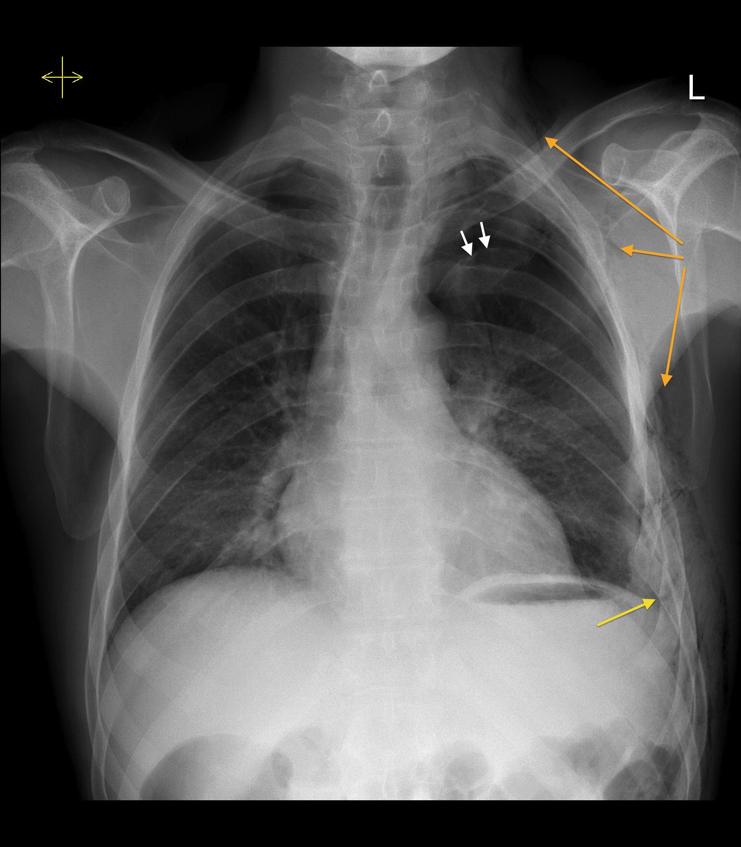
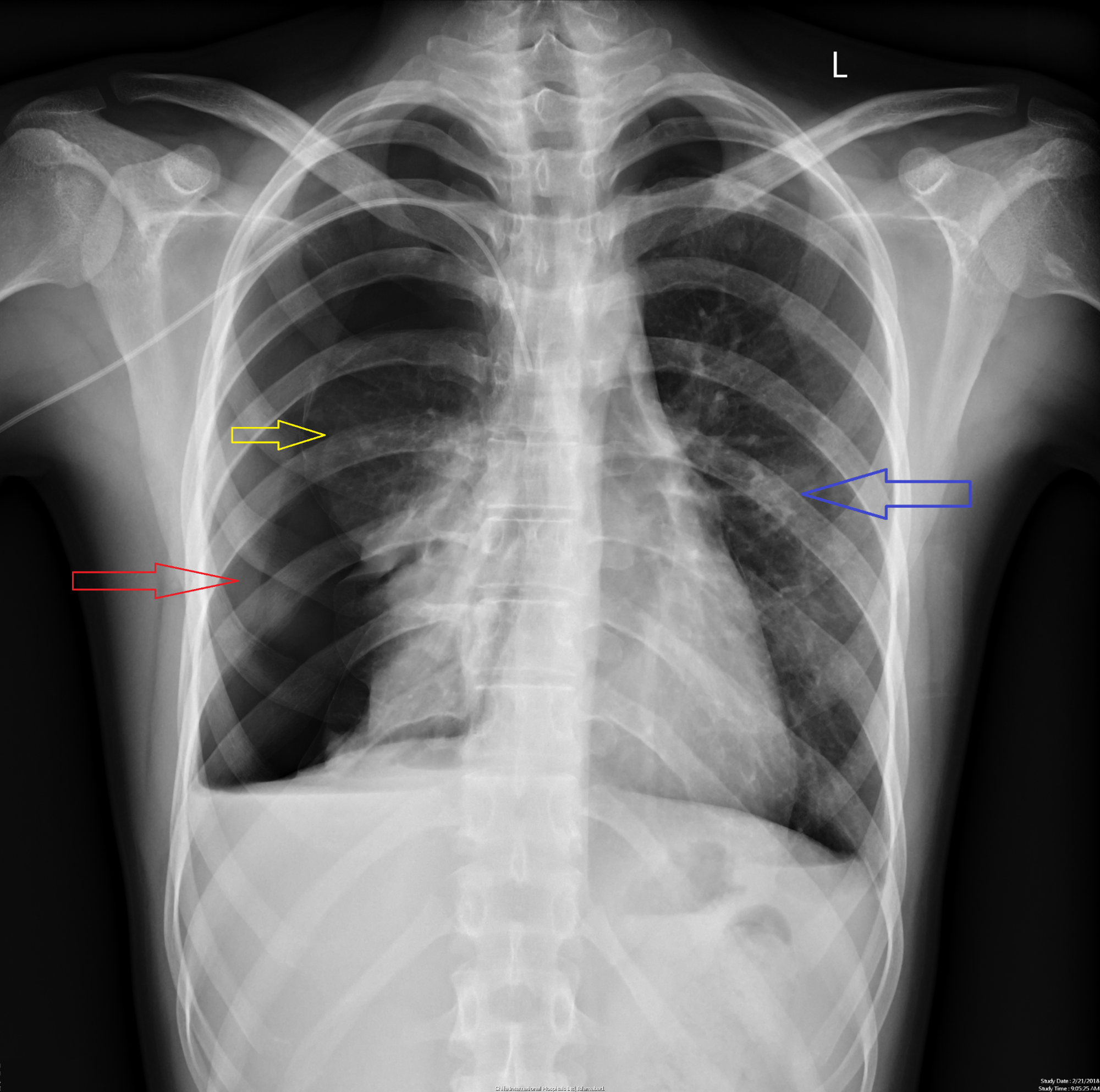
An erect chest radiograph has a sensitivity as high as 92% for detection of a pneumothorax, whilst a supine projection may only detect 50% 6. Instead, the pneumothorax may be demonstrated by looking for the following signs: relative lucency of the involved hemithorax. deep, sometimes tongue-like, costophrenic sulcus: deep sulcus sign 2.
Pneumothorax Hello Doktor
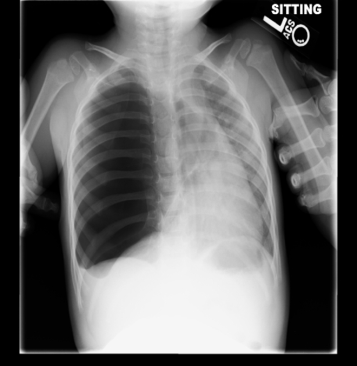
Figure 9.19A AP Chest x-ray, Pneumothorax, Small. Figure 9.19B Lateral Chest x-ray, Pneumothorax, Small. Image Assessment. Findings: The left lung was mildly hyperinflated. There was a visible pleural line in the apex of the left hemithorax. This line was convex outward. There were no visible lung markings beyond this pleural line. Diagnosis:
X Ray Diagnostics Of Pneumothorax Radiographic Representations Of The Lung Tissue In This
Traumatic pneumothorax must be a suspected diagnosis in any blunt or penetrating chest trauma. Adequate history, physical exam and chest X-rays are the mainstays of the diagnosis. However, small pneumothoraces are often missed on physical exam and chest X-ray and may be present on CT chest during a diagnostic workup for other injuries.
'pneumothorax, Xray' Photograph by Du Cane Medical Imaging Ltd
Chest X-ray to tell whether there is air outside the lung; Arterial blood gases; Treatment. Small pneumothoraces may go away on their own. For larger pneumothoraces, the air must be removed from around the lung. A chest tube placed between the ribs into the space around the lungs helps drain the air and allows the lung to re-expand.
Tension pneumothorax due to rib fracture Radiology at St. Vincent's University Hospital
In this video, you'll learn to identify when radiological pleura is abnormal and the key signs to look out for when trying to diagnose a pneumothorax. Want to master chest x-ray interpretation? Take our Chest X-ray Essentials course and learn how to interpret chest x-rays like a pro. Your instructor, Dr Julian Dobrowski-an award-winning.
Pneumothorax Causes, Signs, Symptoms, Treatment
Unlike in pneumothorax, the inner margins of bullae or cysts usually are concave rather than convex and do not conform exactly to the contours of the costophrenic sulcus. A pneumothorax with a pleural adhesion also may simulate bullae or lung cysts.. American Roentgen Ray Society, Canadian Association of Radiologists, Canadian Medical.
Cureus Simultaneous Bilateral Spontaneous Pneumothorax A Rare Complication of
Key Points. Pneumothorax is air in the pleural space causing partial or complete lung collapse. Pneumothorax can occur spontaneously or result from trauma or medical procedures. Diagnosis is based on clinical criteria and chest x-ray. Most pneumothoraces require transcatheter aspiration or tube thoracostomy.
Study Medical Photos Primary Spontaneous Pneumothorax Chest X ray
A large pneumothorax is radiographically defined as one with > 2 cm from pleural surface to lung edge; this is an objective indication for drainage. chest drain bottle is not placed on the trolley above the level of the patient's thorax during the trip to the x ray department. This may result in accumulation of air and fluid in the pleural.
Pneumothorax Radiology For Beginners by Dr. med. Samuel Kobba
Citation, DOI, disclosures and article data. Getting a film with a pneumothorax in the exam is one of the many exam set-pieces that can be prepared for. It is unlikely that they will give you a simple pneumothorax - so, it is worthwhile considering the likely causes and whether it is under tension. Miss it at your peril (both in real life and.
Pneumothorax Concise Medical Knowledge
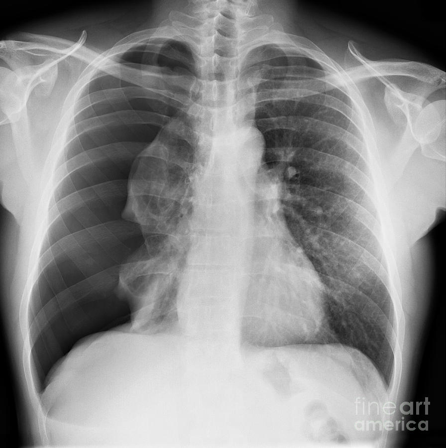
A pneumothorax is generally diagnosed using a chest X-ray. In some cases, a computerized tomography (CT) scan may be needed to provide more-detailed images.. your doctor may simply monitor your condition with a series of chest X-rays until the excess air is completely absorbed and your lung has re-expanded. This may take several weeks.

Image
erect chest x-ray. will show most pneumothoraces; CT chest. will show tiny pneumothoraces not shown on chest x-ray. these are often incidental and asymptomatic; not used for assessment of pneumothoraces unless complex; Radiographic features Plain radiograph. A pneumothorax is seen as a region of lucency (dark) around the edge of the lung.